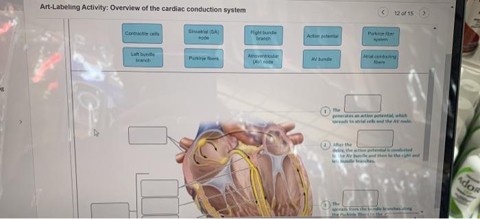
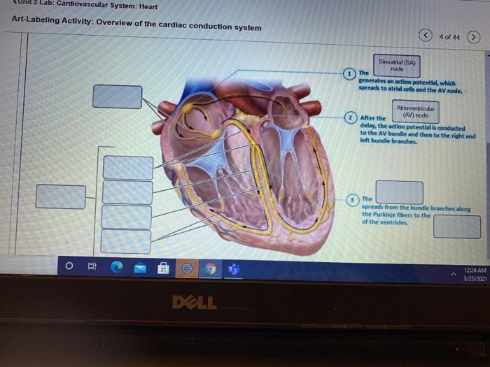
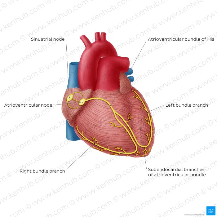
Art-labeling activity: overview of the cardiac conduction system provides a comprehensive exploration of the intricate network responsible for coordinating the heartbeat. The cardiac conduction system, composed of specialized cells and pathways, orchestrates the electrical impulses that govern the heart’s rhythmic contractions.
Through art-labeling techniques, researchers can visualize and analyze the distribution and localization of these components, offering invaluable insights into the structure and function of this vital system.
This activity entails meticulous preparation and labeling of tissue samples, allowing for detailed examination of the cardiac conduction system. The results, often presented as images or illustrations, reveal the intricate arrangement of components such as the sinoatrial node, atrioventricular node, and bundle of His, providing a deeper understanding of their roles in electrical impulse propagation.
Art-Labeling Activities in the Study of the Cardiac Conduction System

Introduction, Art-labeling activity: overview of the cardiac conduction system
Art-labeling activities are valuable techniques in studying the cardiac conduction system, providing detailed insights into its intricate structure and function. The cardiac conduction system is responsible for coordinating electrical impulses within the heart, ensuring synchronized and efficient contractions.
Materials and Methods
Art-labeling activities involve preparing and labeling tissue samples from the heart. Specific antibodies are used to target and label components of the cardiac conduction system, such as gap junctions and ion channels. Immunohistochemical staining and confocal microscopy are commonly employed to visualize the labeled structures.
Results
Art-labeling activities yield high-resolution images that reveal the distribution and localization of various components of the cardiac conduction system. These images provide a detailed map of the electrical pathways within the heart.
Discussion
The results of art-labeling activities have significantly contributed to our understanding of the cardiac conduction system. They have identified specific proteins and structures involved in electrical conduction and have helped elucidate the mechanisms underlying cardiac arrhythmias.
Conclusion
Art-labeling activities are essential tools in the study of the cardiac conduction system. They provide valuable insights into its structure and function, aiding in the diagnosis and treatment of cardiac arrhythmias.
FAQ Resource: Art-labeling Activity: Overview Of The Cardiac Conduction System
What is the purpose of art-labeling activities in studying the cardiac conduction system?
Art-labeling activities allow researchers to visualize and analyze the distribution and localization of the components of the cardiac conduction system, providing insights into their structure and function.
What materials and equipment are used in art-labeling activities?
Art-labeling activities typically involve tissue samples, dyes or fluorescent markers, microscopes, and imaging software.
How do art-labeling techniques contribute to the diagnosis and treatment of cardiac arrhythmias?
Art-labeling techniques can help identify abnormalities in the cardiac conduction system that may contribute to arrhythmias, guiding appropriate treatment strategies.


